Scientific dossier · Hand · Fibroproliferative disease
Dupuytren's Contracture
Also known as Dupuytren's disease or palmar fibromatosis, Dupuytren's contracture is a slowly progressive fibrosis of the palmar aponeurosis. This dossier reviews symptoms, causes, stages, diagnosis and validated treatments — from observation to needle aponeurotomy, collagenase and hand surgery — with a critical look at the scientific evidence.
Introduction
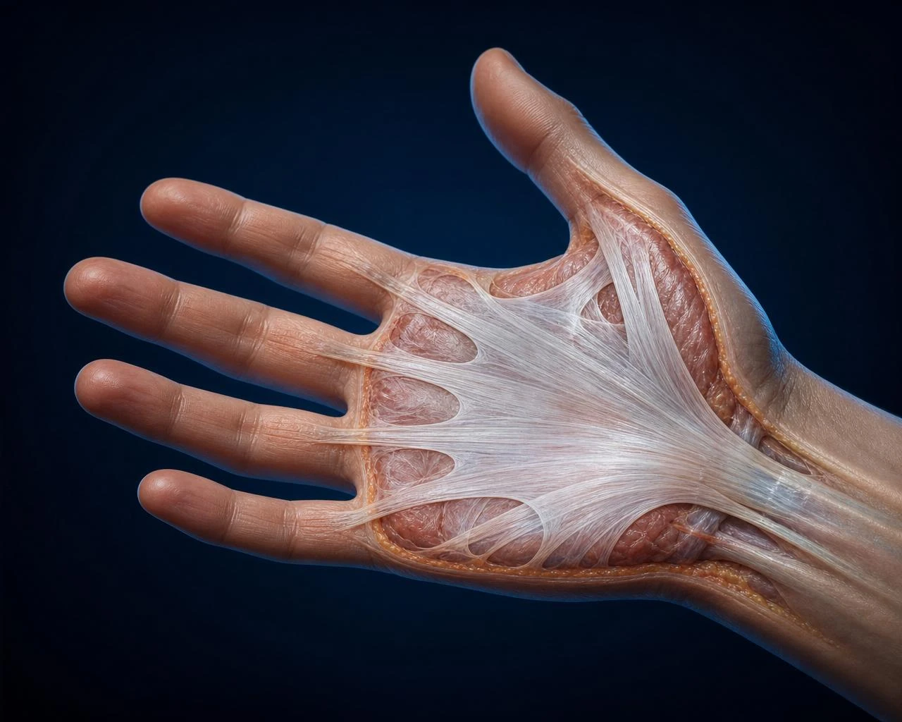
Dupuytren's contracture, also called Dupuytren's disease or palmar fibromatosis, is a benign but progressive fibrosis of the palmar aponeurosis — the fibrous sheet that lies just under the skin of the palm. Firm nodules develop, evolve into taut cords and can eventually bend one or more fingers into a fixed flexion that no longer straightens.
It is most common in adults of Northern European ancestry over the age of 50, more frequent in men, and has a strong hereditary component. The condition itself is not dangerous and does not shorten life expectancy, but advanced forms can significantly impair daily activities such as shaking hands, wearing gloves, washing or reaching into pockets.
This dossier is meant for patients, caregivers and healthcare professionals looking for a clear, neutral, evidence-based summary of the disease, its diagnosis and its validated treatments.
Key takeaways
- Dupuytren's contracture is a slowly progressive fibrosis of the palmar aponeurosis (palmar fascia), also called Dupuytren's disease or palmar fibromatosis.
- It typically starts as a firm palm nodule, evolves into a fibrous cord and can bend one or more fingers permanently — most often the ring and little fingers.
- The exact cause is unknown; genetics, male sex, age > 50, Northern European ancestry, diabetes, tobacco and alcohol are recognised risk factors.
- Diagnosis is clinical. The tabletop test (inability to lay the hand flat on a table) is a simple, reliable red flag.
- Validated treatments include observation, needle aponeurotomy, collagenase injections and open surgery (limited fasciectomy). No dietary supplement is a validated treatment.
Quick facts
| Also known as | Dupuytren's disease, palmar fibromatosis, Viking disease |
| ICD-10 | M72.0 — Palmar fascial fibromatosis |
| Prevalence | 4–6% of adults in Northern Europe; increases sharply after age 50 |
| Sex ratio (M:F) | ~5–10 : 1 (male predominance) |
| Most affected fingers | 4th (ring) and 5th (little) |
| Family history | Present in roughly 60% of patients |
| Typical course | Slow, progressive; years to decades |
| Reference specialty | Hand surgery / orthopaedics |
Did you know?
4–6%
Prevalence
Adults in Northern Europe; increases with age.
Did you know?
> 50 yrs
Peak onset
Rare before age 40.
Did you know?
× 5–10
Sex ratio (M:F)
Marked male predominance.
Did you know?
4th – 5th
Most affected fingers
Ring and little fingers first.
Did you know?
≈ 60%
Family history
Strong genetic component suspected.
What is Dupuytren's contracture?
Dupuytren's contracture is a fibroproliferative disorder of the palmar fascia. Fibroblasts in the aponeurosis differentiate into contractile myofibroblasts that produce excess type-III collagen and actively pull on the tissue. Over time, this remodelling forms palpable nodules and longitudinal cords that shorten the aponeurosis and bend the fingers.
Histologically, the disease evolves through three phases described by Luck: a proliferative phase (myofibroblast expansion), an involutional phase (dense collagen deposition and active contraction) and a residual phase (mature, acellular fibrous cords). Clinically, these phases roughly map to nodule, cord and fixed contracture.
It belongs to the same family as Ledderhose disease (plantar fibromatosis) and Peyronie's disease (fibromatosis of the tunica albuginea) and can coexist with them.
Healthy hand
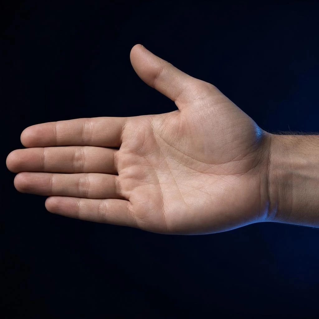
Supple aponeurosis · full finger extension
Affected hand
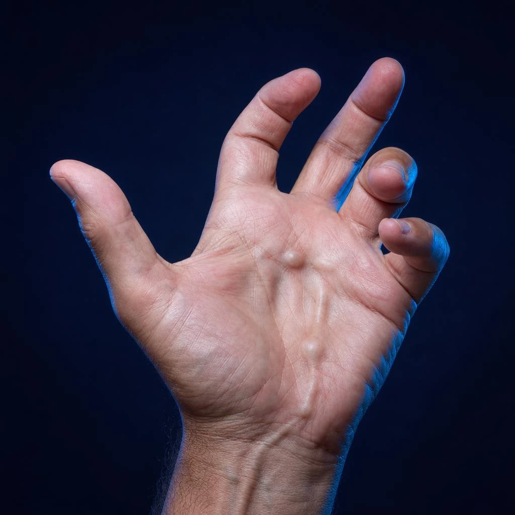
Nodule, fibrous cord, finger contracture
Symptoms
Symptoms usually appear gradually, without pain, and progress over months to years. The three cardinal features are palm nodules, fibrous cords and finger contracture.
Palm nodule
Firm, well-defined lump, most often near the base of the ring finger. Usually painless. May have overlying skin dimpling.
Fibrous cord
Taut band running from the palm toward one or more fingers. Becomes more visible when the fingers are extended.
Finger contracture
Progressive, fixed flexion of the finger. Extension becomes limited, then impossible without treatment.
Positive tabletop test
Inability to lay the palm and fingers flat on a table — a simple screening sign for advanced disease.
Palm nodule
Firm, often painless lump in the palm — the first clinical sign.
Fibrous cord
Longitudinal palpable band under the palm skin extending toward the finger.
Finger contracture
Irreducible flexion: the finger no longer straightens fully — functional impact.
Early-stage Dupuytren's contracture
Early-stage Dupuytren's contracture is defined by a palm nodule or mild skin thickening without a fixed finger contracture. At this stage, the fingers still extend fully and daily activities are not impaired.
Detection is often incidental — a partner or a clinician notices a firm lump in the palm. Skin pitting or dimpling is a subtle but characteristic clue. Most early nodules progress slowly; a minority remain stable for years or even indefinitely.
| Criterion | Early stage | Advanced disease |
|---|---|---|
| Palm changes | One or more firm, painless nodules — often near the base of the ring finger. | Well-formed longitudinal cord extending toward one or several fingers. |
| Skin | Normal or mild pitting/dimpling of the palm skin. | Marked puckering, thickened plaques adherent to underlying fascia. |
| Finger position | Full extension preserved; tabletop test negative. | Fixed flexion of MCP and/or PIP joints; tabletop test positive. |
| Function | Little or no functional impact. | Difficulty shaking hands, wearing gloves, washing face, reaching into pockets. |
| Typical management | Observation, education, hand-therapy advice. | Needle aponeurotomy, collagenase injections or surgery. |
What does Dupuytren's contracture look like?
Dupuytren's contracture typically appears as a firm, painless lump on the palm — most often at the base of the ring finger — sometimes with dimpling of the overlying skin. As the disease progresses, a rope-like cord becomes visible running from the palm toward the finger, and the finger itself becomes progressively bent, unable to be fully straightened.
In advanced disease, the affected fingers are held in permanent flexion, with the fingertips curling toward the palm. The palm skin often looks puckered and thickened. In very severe forms, hygiene of the palm becomes difficult and skin maceration can occur.
Which fingers are affected?
Any finger can be affected, but the ring finger (4th) is the most common, followed by the little finger (5th) and the middle finger (3rd). The thumb and index finger are involved less often, generally in more aggressive forms.
Bilateral involvement is frequent; disease often appears in the second hand within a few years of the first. Involvement of multiple fingers on both hands, especially in a younger patient, defines a Dupuytren diathesis: a more aggressive phenotype with higher recurrence after treatment.
What causes Dupuytren's contracture?
The exact cause of Dupuytren's contracture is unknown. It results from the interaction between a strong genetic predisposition and several acquired risk factors. At the tissue level, the trigger is an abnormal fibroproliferative response of palm fibroblasts to unknown local signals.
Genetic studies (GWAS) have identified variants in the Wnt signalling pathway associated with Dupuytren's disease. Fibroblasts differentiate into myofibroblasts expressing α-smooth muscle actin, driven by TGF-β1, TNF and oxidative stress signals. These cells actively contract and deposit excess collagen, creating the palpable cord.
Genetics
Wnt pathway variants, familial clustering.
Cellular biology
Myofibroblast differentiation driven by TGF-β1 and TNF.
Oxidative stress
Local ROS production sustains fibroproliferation.
Risk factors
Several factors independently increase the risk of developing Dupuytren's contracture. Most are non-modifiable (genetics, sex, age), but a few lifestyle factors (tobacco, alcohol, diabetes control) can be acted on.
| Factor | Effect | Strength |
|---|---|---|
| Northern European ancestry | Higher prevalence (Viking disease). Genetic component involving Wnt signalling pathways. | Strong |
| Male sex | 5–10× higher risk than women; earlier onset and more severe course. | Strong |
| Age > 50 years | Prevalence rises sharply with age; rare before 40. | Strong |
| Family history | Autosomal-dominant pattern with variable penetrance in many families. | Strong |
| Diabetes mellitus | Consistently associated; often milder, more distal disease. | Moderate |
| Tobacco smoking | Associated with increased incidence and recurrence, likely via microvascular damage. | Moderate |
| Excess alcohol | Associated with higher prevalence, independent of liver disease. | Moderate |
| Manual labour / trauma | Historical hypothesis; current evidence is inconsistent. | Suggestive |
| Epilepsy / antiepileptic drugs | Older reports of association; mechanism unclear. | Suggestive |
Disease progression
Dupuytren's contracture progresses in an unpredictable, non-linear way. Many patients remain at the nodule stage for years or decades, while others develop finger contractures within months. Four stages describe the typical clinical trajectory.
- Stage 1
Palm nodule
Firm painless lump; extension preserved; tabletop test negative.
- Stage 2
Skin changes and early cord
Skin dimpling, pretendinous band appearing; mild MCP flexion possible.
- Stage 3
Established contracture
Well-formed cord; fixed MCP and/or PIP flexion; tabletop test positive.
- Stage 4
Severe contracture
Marked flexion of one or more fingers; significant functional impairment.
- Stage 0
Normal hand
No palpable abnormality; full finger extension.
- Stage 1
Nodule
Small, usually painless palm lump — first clinical sign.
- Stage 2
Fibrous cord
Longitudinal palpable band running toward the finger.
- Stage 3
Finger contracture
Irreducible flexion; active extension limited.
- Stage 4
Severe contracture
Finger locked in flexion; marked functional impact.
Visual progression
The four anatomical stages of Dupuytren's contracture
From a healthy aponeurosis to severe finger contracture — medical illustrations and biological correlates.
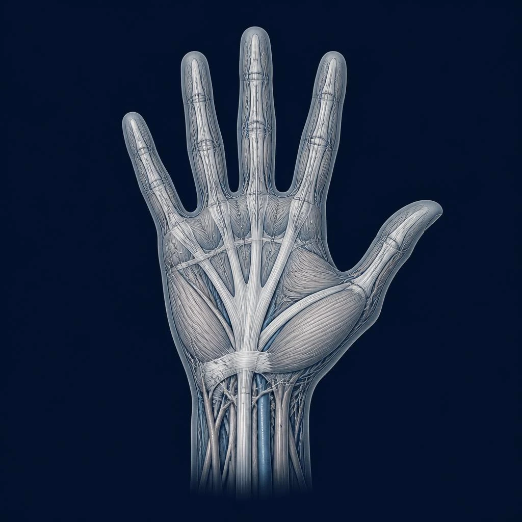 Stage 01
Healthy aponeurosis
Normal hand
Clinical signs
The palm is supple and painless, with no palpable nodule. Fingers extend fully.
Biological correlate
Quiescent fibroblasts. Physiological turnover of type I collagen within the palmar aponeurosis.
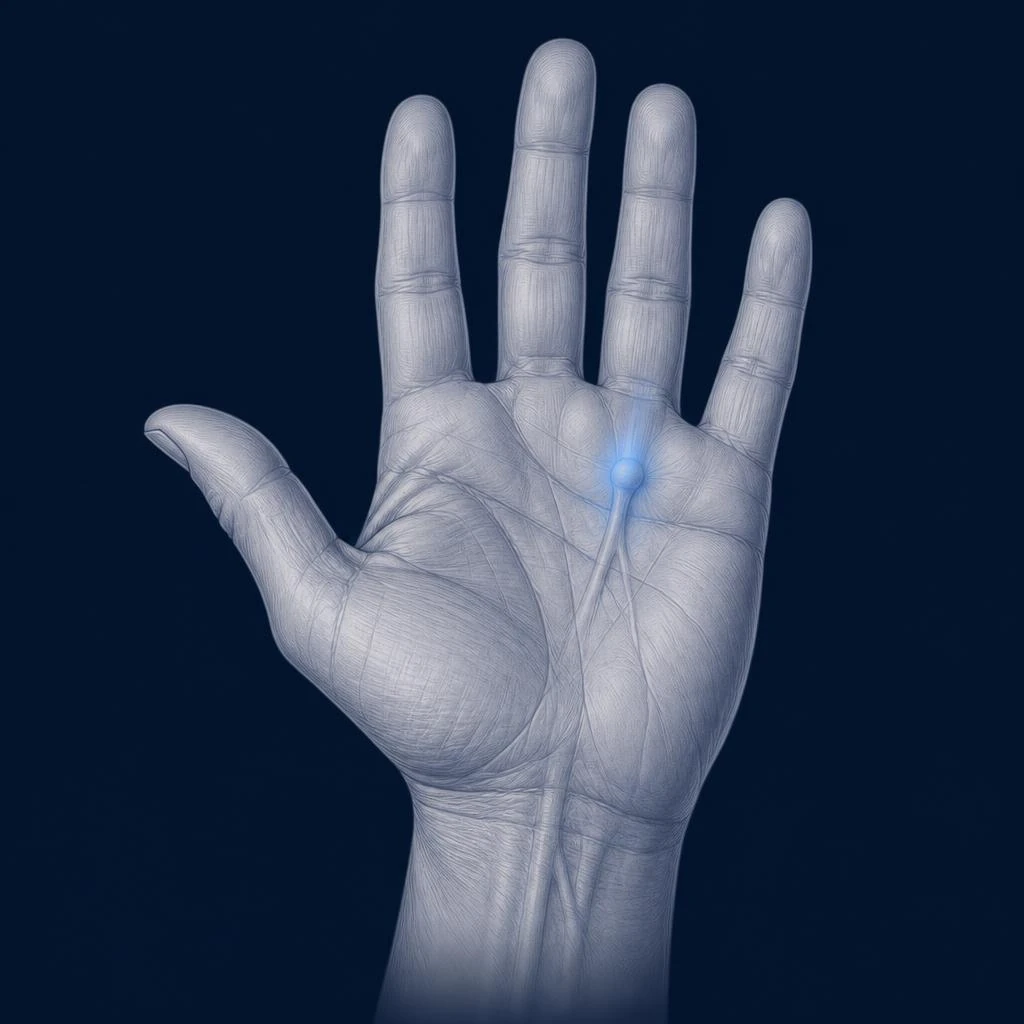 Stage 02
First clinical sign
Palm nodule
Clinical signs
Firm, painless nodule most often over the 4th ray. Finger extension still preserved.
Biological correlate
Fibroblast proliferation and differentiation into α-SMA-expressing myofibroblasts. Increased TGF-β1 secretion.
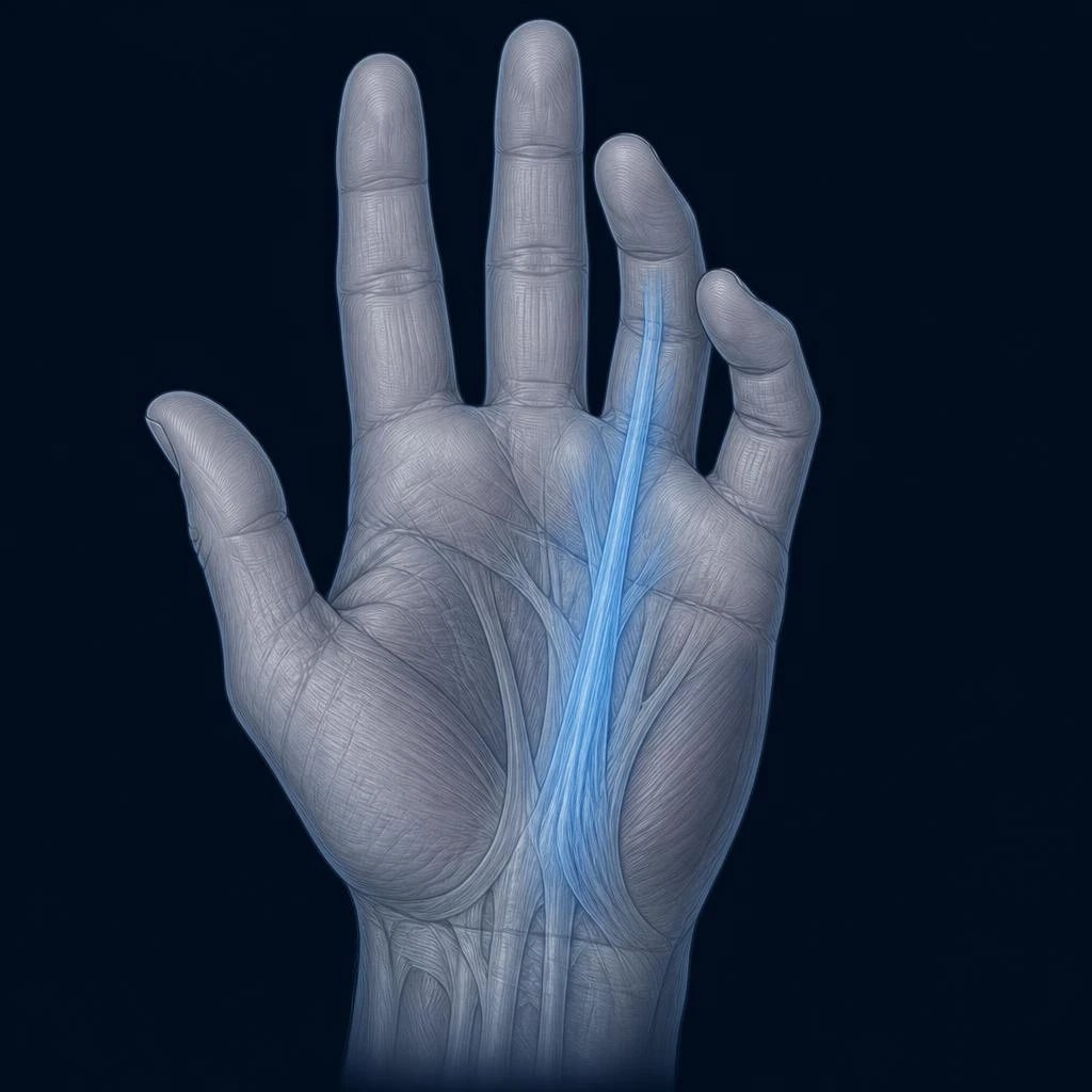 Stage 03
Early contracture
Fibrous cord
Clinical signs
Palpable longitudinal cord from wrist to affected finger. Moderate flexion of the 4th finger (~30°); tabletop test positive.
Biological correlate
Dense collagen matrix organisation (collagen I ↑, MMP/TIMP imbalance). Active myofibroblast contraction.
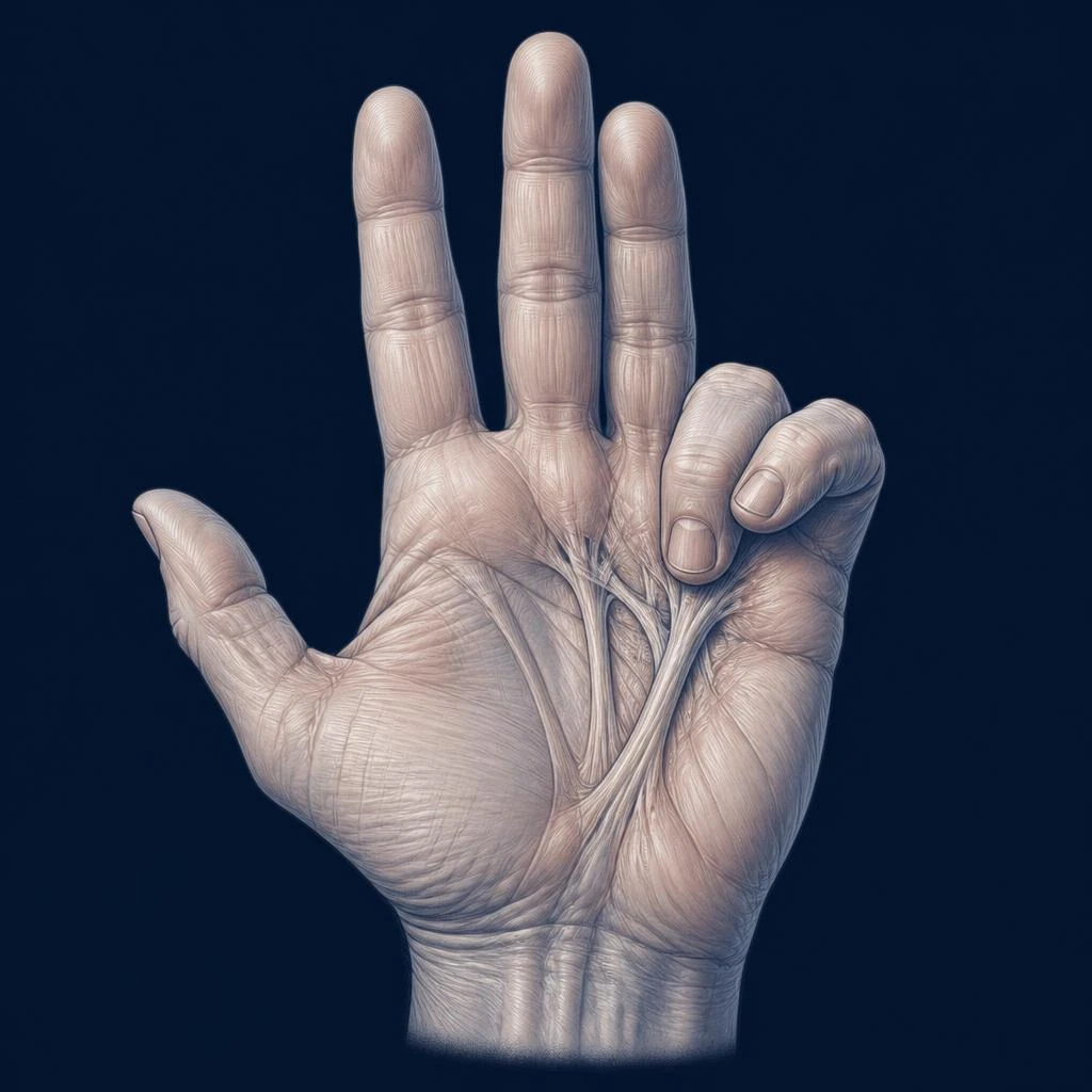 Stage 04
Disabling contracture
Severe contracture
Clinical signs
Permanent flexion of the 4th (~90°) and often 5th finger. Marked functional impairment: surgical indication (fasciectomy) or needle aponeurotomy.
Biological correlate
Persistent mature fibrosis. ROS / NF-κB pathway sustaining local inflammation. Myofibroblast resistance to apoptosis.
Does Dupuytren's contracture affect life expectancy?
Dupuytren's contracture is a benign condition and does not shorten life expectancy on its own. Some cohort studies have reported a modest association with cardiovascular disease and all-cause mortality, but this most likely reflects shared risk factors (age, sex, diabetes, alcohol, tobacco) rather than a direct effect of Dupuytren's disease.
Patients with Dupuytren's contracture should nevertheless benefit from the same cardiometabolic prevention as any adult with similar risk factors: blood-pressure control, diabetes management, smoking cessation and moderation of alcohol.
Dupuytren vs similar conditions
Several hand conditions can be confused with Dupuytren's contracture, especially at an early stage. A hand specialist can differentiate them clinically.
| Condition | Key feature | Distinctive sign |
|---|---|---|
| Dupuytren's contracture | Fibrous cord/nodule in the palmar aponeurosis, progressive finger flexion. | Skin dimpling, tabletop test positive, painless. |
| Trigger finger (stenosing tenosynovitis) | Painful catching/locking of a finger during flexion. | Nodule at the A1 pulley over the flexor tendon; no palmar cord. |
| Ledderhose disease (plantar fibromatosis) | Similar fibromatosis on the sole of the foot. | Often coexists with Dupuytren; same fibroproliferative family. |
| Camptodactyly | Congenital fixed flexion of the little finger. | Present from childhood; no palm nodule or cord. |
| Post-traumatic scar contracture | Fixed flexion after a hand injury or burn. | Clear traumatic history, visible scar, no diffuse aponeurosis involvement. |
Diagnosis
Diagnosis is clinical. The physician takes a history, inspects and palpates the palm and fingers, and quantifies the finger contracture. No imaging or blood test is required in the vast majority of cases.
History
Onset, progression, family history, dominant hand, diabetes, tobacco, alcohol, occupation.
Inspection & palpation
Identify nodules, cords, skin dimpling; assess mobility of each finger joint.
Tabletop test (Hueston)
Ask the patient to lay the hand flat on a table: inability to do so = positive test.
Goniometry
Measure flexion angles at MCP and PIP joints to grade severity and guide treatment.
Clinical observation
Tabletop test
Extension measurement
Functional assessment
When should you see a hand specialist?
You should see a hand surgeon or an orthopaedic hand specialist as soon as a finger contracture appears, the tabletop test becomes positive, or the disease starts to interfere with daily activities. Earlier referral is also appropriate if you have a young age at onset, bilateral disease or a strong family history.
When to seek medical advice
- A finger no longer straightens completely.
- You cannot lay your palm flat on a table (positive tabletop test).
- Daily activities (shaking hands, wearing gloves, washing) become difficult.
- A palm nodule grows quickly or becomes painful.
- Disease appears before age 50 or on both hands with several fingers involved.
Treatment options
There is no single best treatment. Guideline-based care combines observation, hand therapy, needle aponeurotomy, collagenase injections and surgery. Choice depends on the severity, the joints affected, recurrence risk and patient preferences.
| Option | What it is | Best for | Benefits | Limits | Evidence |
|---|---|---|---|---|---|
| Observation | Watchful waiting with periodic reassessment of finger extension. | Isolated palm nodules, no functional impact, negative tabletop test. | No procedure, no downtime; many nodules progress slowly or not at all. | Requires regular follow-up to detect progression. | Strong evidence |
| Hand therapy | Stretching, splinting and functional exercises supervised by a hand therapist. | Post-procedural care; maintaining range of motion. | Improves recovery after aponeurotomy, collagenase or surgery. | No proven ability to reverse an established contracture on its own. | Moderate evidence |
| Needle aponeurotomy (PNA) | Percutaneous division of the cord with a fine needle under local anaesthesia. | MCP contracture, single well-defined cord. | Minimally invasive, immediate improvement, fast return to activity. | Higher recurrence than surgery; less effective on PIP contracture. | Strong evidence |
| Collagenase injections | Injection of Clostridium histolyticum collagenase to enzymatically weaken the cord, followed by cord rupture. | Palpable cord causing MCP contracture; PIP with caution. | Non-surgical, effective on MCP joints in randomised trials. | Bruising, skin tears; not available in all countries; recurrence possible. | Strong evidence |
| Limited fasciectomy (surgery) | Open surgical removal of the diseased fascia. | Severe contracture, PIP involvement, revision after recurrence. | Lowest recurrence rate among validated options. | Longer recovery, wound complications, risk of nerve/vessel injury. | Strong evidence |
| Dermofasciectomy | Fasciectomy combined with skin excision and full-thickness graft. | Aggressive/recurrent disease, marked skin involvement. | May reduce local recurrence in high-risk patients. | More complex procedure; longer immobilisation; scarring. | Moderate evidence |
Needle aponeurotomy vs collagenase vs surgery
The three main procedural treatments have different profiles in terms of invasiveness, recovery, effectiveness and recurrence. This side-by-side comparison summarises the practical differences.
| Criterion | Needle aponeurotomy | Collagenase injection | Limited fasciectomy |
|---|---|---|---|
| Invasiveness | Percutaneous needle | Injection + manipulation | Open surgery |
| Anaesthesia | Local | Local | Local or regional |
| Recovery | Days | 1–2 weeks | Weeks to months |
| Best joint target | MCP | MCP (± PIP) | MCP and PIP |
| Effectiveness (MCP) | High, short-term | High (CORD-I/II) | Highest, most durable |
| Recurrence at 3–5 y | Higher | Moderate | Lowest |
| Main risks | Nerve/tendon injury (rare), skin tear | Bruising, skin tear, oedema | Wound problems, nerve/artery injury, CRPS |
Recurrence
Because Dupuytren's disease is a chronic biological process, recurrence is possible after any treatment. Rates depend on the technique, the joints treated and patient-related factors.
- Needle aponeurotomy: recurrence around 50–65% at 5 years (van Rijssen et al., 2012).
- Collagenase injections: intermediate recurrence, often around 35–50% at 5 years depending on definitions.
- Limited fasciectomy: lowest recurrence, roughly 20–30% at 5 years in most series.
A Dupuytren diathesis (young age, bilateral disease, family history, Ledderhose disease, knuckle pads) is the strongest predictor of recurrence, regardless of the treatment used.
Exercises and hand therapy
Exercises and stretching cannot reverse an established Dupuytren cord. However, structured hand therapy plays a central role after needle aponeurotomy, collagenase injections or surgery, to preserve range of motion, control oedema and prevent stiffness.
A hand therapist typically prescribes gentle extension exercises, oedema-control techniques and, when appropriate, a static extension splint worn at night for several weeks or months. Aggressive or painful stretching should be avoided as it can increase inflammation.
Foods and supplements
No specific food is proven to cause, prevent or cure Dupuytren's contracture. There is no scientifically validated "Dupuytren diet". General health measures — a balanced Mediterranean-style diet, tight diabetes control, moderation of alcohol and avoidance of tobacco — remain reasonable recommendations, mostly for their overall cardiometabolic benefit.
Some patients ask about vitamin E, N-acetylcysteine, curcumin or other antioxidant supplements. Older trials of vitamin E in Dupuytren's disease did not confirm a clinical benefit. Newer molecules are studied only at the preclinical level (see below).
Current research
Research on Dupuytren's contracture focuses on the biology of myofibroblasts, the role of TGF-β1 and TNF, prognostic biomarkers, and new pharmacological approaches aimed at slowing early-stage disease rather than treating advanced contractures.
- Anti-TNF (adalimumab). The RIDD trials (Nanchahal et al., 2018 and 2022) provided randomised evidence that intranodular adalimumab reduces nodule size and softens tissue in early-stage disease. Long-term effects on progression to contracture are still being evaluated.
- Genetics. Genome-wide association studies have confirmed Wnt-pathway involvement, opening the way to targeted therapies (Dolmans et al., 2011).
- Refinements of collagenase and needle aponeurotomy. Ongoing work refines patient selection, technique and post-procedural care to reduce recurrence.
- Antioxidant and anti-fibrotic pathways. Preclinical work on ROS, TGF-β1 and NF-κB continues to inform hypotheses on how nutritional or pharmacological modulation might influence fibrosis.
Cascade · TGF-β → contracture
Shared mechanism · fibroproliferative diseases
Collagen organization · healthy vs fibrosis
Dietary supplements: what the science actually says
Dietary supplements are not a validated treatment for Dupuytren's contracture. Some molecules — antioxidants, polyphenols — are studied for their potential role in oxidative stress, inflammation and fibrosis pathways, but clinical evidence remains limited or absent. They cannot, on their own, prevent, halt or cure the disease.
For a detailed cross-cutting analysis of nutritional approaches studied in fibroproliferative diseases and their levels of evidence, see our dossier on natural approaches and fibroproliferative diseases.
| Ingredient | Proposed biological mechanism | Quality of evidence |
|---|---|---|
| Curcumin | Modulation of TGF-β1 and NF-κB signalling in preclinical fibrosis models. | Preclinical only |
| Quercetin | Antioxidant flavonoid; senolytic activity on myofibroblasts in cell studies. | Preclinical only |
| Resveratrol | Modulates ROS and TGF-β1; explored in fibroproliferative cell models. | Preclinical only |
| Ginkgo biloba | Antioxidant flavone glycosides; microcirculation support. | Limited evidence |
| Pomegranate polyphenols | Anti-inflammatory and antioxidant properties studied in vitro. | Preclinical only |
| Vitamin E (α-tocopherol) | Historical trials in Dupuytren; no confirmed clinical benefit. | Limited evidence |
Current clinical evidence for these compounds as a treatment of Dupuytren's contracture is very limited. Dietary supplements do not replace medical care and should be discussed with a healthcare professional before use.
Frequently asked questions
Scientific references
A selection of reference publications and international guidelines used to write this dossier. The list is not exhaustive and will be enriched as the dossier is updated.
- [1]Hindocha S et al., 2006 (). Dupuytren's diathesis revisited: evaluation of prognostic indicators for risk of disease recurrence. · Voir la source
- [2]Hurst LC et al., 2009 (CORD I) (). Injectable collagenase clostridium histolyticum for Dupuytren's contracture. · Voir la source
- [3]Gilpin D et al., 2010 (CORD II) (). Injectable collagenase Clostridium histolyticum: a new nonsurgical treatment for Dupuytren's disease. · Voir la source
- [4]van Rijssen AL et al., 2012 (). Five-year results of a randomized clinical trial on treatment in Dupuytren's disease: percutaneous needle fasciotomy versus limited fasciectomy. · Voir la source
- [5]Werker PMN et al., 2012 (). Percutaneous needle fasciotomy for Dupuytren's disease. · Voir la source
- [6]Verjee LS et al., 2013 (). Unraveling the signaling pathways promoting fibrosis in Dupuytren's disease reveals TNF as a therapeutic target. · Voir la source
- [7]Ball C et al., 2016 (Cochrane) (). Interventions for treating Dupuytren's contracture. · Voir la source
- [8]Larsen S et al., 2015 (). Genetic and environmental influences in Dupuytren's disease: a twin study. · Voir la source
- [10]Nanchahal J et al., 2018 (RIDD) (). Anti-TNF therapy for Dupuytren's disease: a randomised dose-response proof-of-concept trial. · Voir la source
- [11]Nanchahal J et al., 2022 (RIDD phase 2b) (). Adalimumab for early-stage Dupuytren's disease: a multicentre, randomised, placebo-controlled trial. · Voir la source
- [13]American Society for Surgery of the Hand (ASSH) (). Dupuytren's disease patient information. · Voir la source
- [14]Cochrane Library (). Systematic reviews — Dupuytren's contracture interventions. · Voir la source
Conclusion
Dupuytren's contracture is a common, benign but progressive fibrosis of the palmar aponeurosis. Modern care combines observation of early disease with minimally invasive procedures (needle aponeurotomy, collagenase) and, when necessary, hand surgery, all tailored to the joints affected, the severity of the contracture and the patient's expectations.
AARO LAB focuses on the biological mechanisms involved in fibrosis, oxidative stress and tissue integrity. Dietary supplements are not a treatment for Dupuytren's disease and cannot replace an evaluation by a hand specialist.
Related reading
Fibrosis
Myofibroblasts, TGF-β1 and collagen remodelling across organs.
Oxidative stress
ROS, redox balance and their role in fibroproliferative diseases.
Extracellular matrix
Structure, remodelling and pathology of the ECM.
Peyronie's disease
The same fibroproliferative family, applied to the tunica albuginea.
Natural approaches — fibroses
Critical reading of ingredients studied in Peyronie and Dupuytren.
Institutional sources
Further reading
These institutional resources let you explore the topic further via authoritative scientific or medical organisations.
- PubMedPubMed / National Library of MedicineDupuytren's disease — clinical researchCorpus scientifique sur la physiopathologie et la prise en charge de la maladie de Dupuytren.
- CochraneCochrane LibraryInterventions for Dupuytren's contractureRevues systématiques comparant chirurgie, aponévrotomie et collagénase.
- HASHaute Autorité de SantéRecommandations HAS — chirurgie de la mainRecommandations françaises applicables à la chirurgie de la main et à Dupuytren.
- NIHNational Institutes of HealthNIH — Dupuytren's disease overviewFiche scientifique de l'Institut national américain sur les pathologies rhumatologiques.
External links to scientific or medical organisations. AARO LAB has no commercial ties with the sites cited.