Peyronie's Disease
Peyronie's disease is a benign fibrotic disorder of the tunica albuginea of the penis that causes an acquired penile curvature during erection, often associated with pain, penile shortening and, in some cases, erectile dysfunction. This evidence-based dossier explains the symptoms, causes, stages, diagnosis, treatment options and current research, drawing on EAU 2024 and AUA 2015/2021 guidelines and the PubMed-indexed literature.
Introduction
Peyronie's disease (induratio penis plastica) is a benign but often disabling connective-tissue disorder of the erectile envelope of the penis (the tunica albuginea). First described by François Gigot de La Peyronie in 1743, it is characterised by the formation of a fibrous plaque that impairs the extensibility of the tunica and leads to an acquired penile curvature.
Contemporary epidemiological studies estimate its prevalence between 3 and 9% of the adult male population, with a marked increase after 50 years of age and in men with diabetes. It is therefore far more common than once thought, which justifies better public information and specialised care.
Key takeaways
- Peyronie's disease is a benign fibrotic disorder of the tunica albuginea that causes an acquired penile curvature during erection.
- It typically evolves in two phases: an active (inflammatory) phase of 6–18 months and a stable (chronic) phase, when the plaque and curvature no longer progress.
- There is no universal cure. Guideline-based care (EAU, AUA) combines observation, intralesional injections, penile traction, vacuum devices and, when needed, corrective surgery.
- Dietary supplements are not a validated treatment for Peyronie's disease. Some molecules (antioxidants, polyphenols) are studied for their role in oxidative stress and fibrosis pathways, with limited clinical evidence.
- Early urological consultation improves diagnostic accuracy and expands the range of options available during the active phase.
Prevalence in adult men
≈ 3–9%
Peak age
50–60 years
Typical active phase
6–18 months
Total course to stabilisation
12–24 months
Guidelines
EAU 2024 · AUA 2015/2021
Main tissue involved
Tunica albuginea
Key figures · at a glance
Did you know?
3–9 %
Estimated prevalence
Adult men according to contemporary studies.
Did you know?
50–60
Mean age (years)
Peak incidence reported in the literature.
Did you know?
~70 %
Pain in the active phase
Mainly during erection.
Did you know?
~30–50 %
Associated erectile dysfunction
Varies by series and severity.
Did you know?
12–18 months
Inflammatory phase
Average duration before stabilisation.
What is Peyronie's disease?
Peyronie's disease is an acquired disorder in which a fibrous plaque forms inside the tunica albuginea of the penis. Because this plaque is stiffer than the surrounding tissue, the penis bends toward it during erection, producing a curvature that may be associated with pain, shortening or erectile dysfunction.
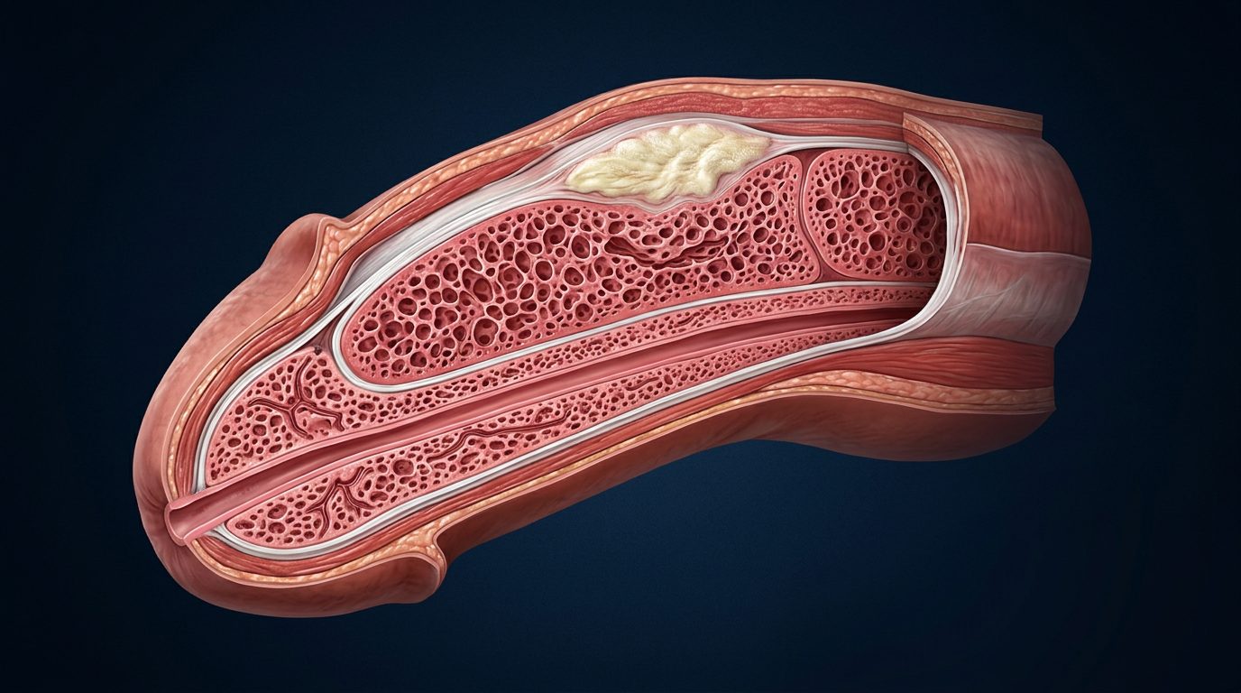
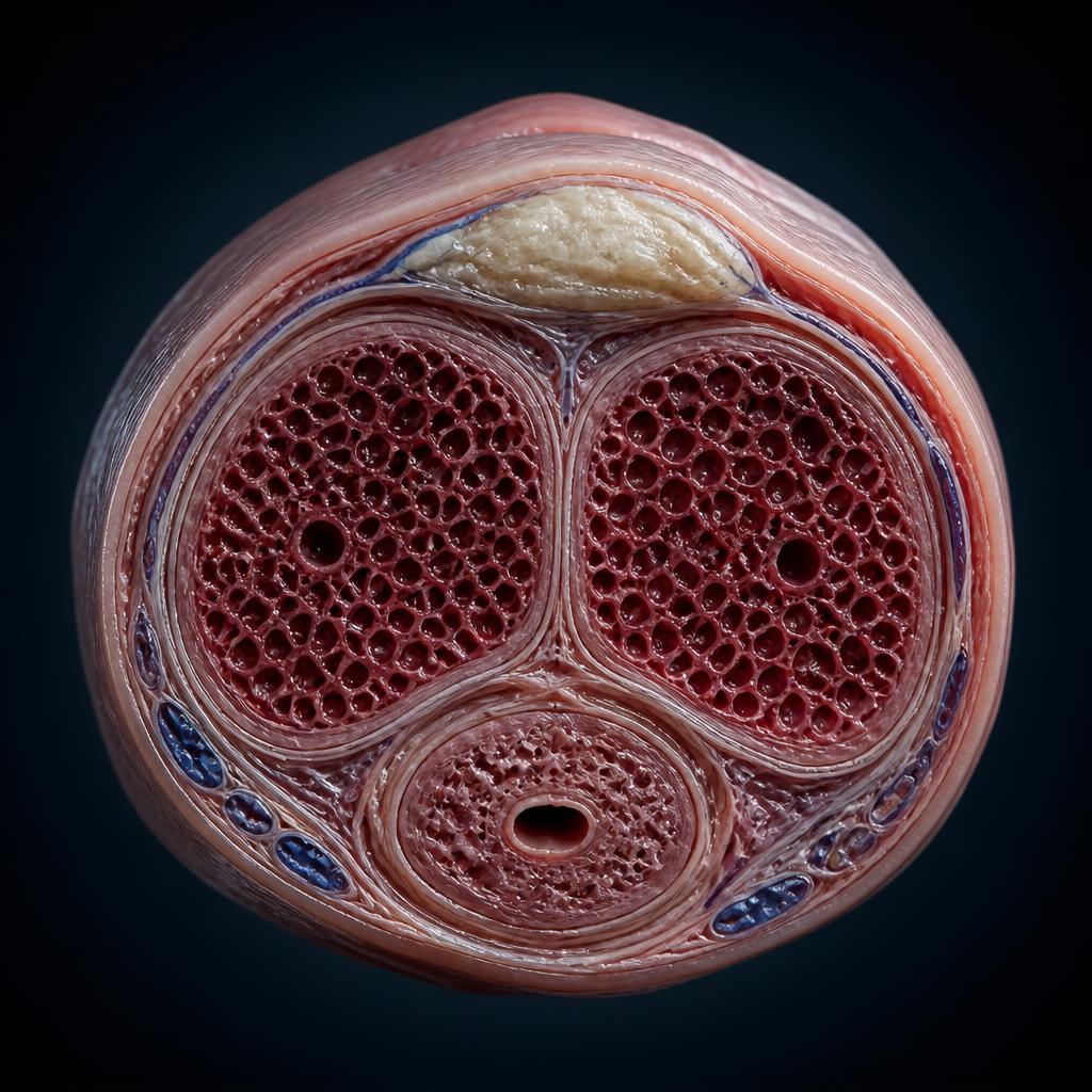
To understand the disease, one first needs the anatomy. The penis contains two corpora cavernosa (responsible for erection) and a corpus spongiosum (around the urethra). The corpora cavernosa are wrapped in a resistant fibrous membrane — the tunica albuginea — primarily made of type I and III collagen. By distending uniformly during erection, this envelope gives the penis its rigidity.
In Peyronie's disease, a fibrous plaque develops within the tunica. Being stiffer than normal tissue, it loses the ability to extend during erection: as the opposite side continues to stretch normally, the penis deforms into a curvature. The plaque may also calcify and become palpable as an induration under the skin.
Peyronie's plaque is a localised form of penile-tissue fibrosis. For a detailed analysis at the level of the tunica and cavernous bodies, see our dedicated dossier on penile fibrosis.
Longitudinal anatomy
Transverse cross-section
Symptoms
The most common symptoms of Peyronie's disease are penile curvature during erection, penile pain (mainly in the active phase), a palpable plaque on the shaft, penile shortening and, in some patients, erectile dysfunction. Severity varies widely from one man to another.
Penile curvature
Acquired deviation appearing during erection — dorsal, ventral or lateral — of variable angle, sometimes with an hourglass narrowing.
Penile pain
Present mainly during the active phase, on erection or occasionally at rest; usually regresses spontaneously as the disease stabilises.
Penile shortening
Loss of erect length is reported by a significant proportion of patients.
Erectile dysfunction
Insufficient rigidity may coexist, related to tissue involvement and/or associated vascular factors.
Palpable plaque
Indurated area felt on palpation, most often dorsal, sometimes multiple.
Psychological distress
Anxiety, reduced self-esteem and impact on the couple are frequently reported.
Early signs to recognise
Early signs of Peyronie's disease can be subtle. Recognising them early makes it possible to start care during the active phase, when several treatments are most effective.
Emerging penile pain
A painful sensation on erection or at rest, often the first sign reported, appearing weeks before the curvature becomes obvious.
Palpable nodule or plaque
Small induration felt under the skin of the penis, of recent onset, usually painless or only mildly tender on palpation.
Progressive curvature
A mild deviation that increases over several weeks or months during the active phase.
Loss of length
Sensation of a shorter penis on erection, sometimes preceding an obvious curvature.
Erectile difficulties
Reduced rigidity, difficulty maintaining erection or instability of the erection distal to the plaque.
When to consult
As soon as any of these signs appear. Early urological consultation improves diagnostic accuracy and treatment options.
Penile pain: what to know
Pain is one of the first signs reported by patients, occurring mainly during the active phase, on erection, and occasionally at rest. Intensity varies from mild discomfort to a level marked enough to cause sexual avoidance. Importantly, pain does not reflect the severity of the curvature: some patients have a pronounced curvature without pain, while others suffer more with a moderate curvature.
In the vast majority of cases, pain regresses spontaneously as the disease enters the stable phase, which can take several months. Persistence of pain beyond 18 months is less common. If pain significantly affects quality of life, a urologist can propose specific management, especially during the active phase.
What causes Peyronie's disease?
Peyronie's disease is thought to result from an abnormal wound-healing response after repeated microtrauma of the tunica albuginea, most often during intercourse. Chronic inflammation, oxidative stress, a genetic background and vascular or metabolic comorbidities appear to amplify the fibrotic response. No single cause has been demonstrated to date.
Microtrauma
Repeated microtrauma of the tunica albuginea during intercourse is the most widely accepted aetiological hypothesis (EAU Guidelines).
Chronic inflammation
A prolonged inflammatory response is thought to promote collagen deposition and the formation of a fibrous plaque.
Oxidative stress
Increased ROS production within the injured tunical tissue is described as a mechanism involved in local fibrosis.
Genetic predisposition
A genetic background is suspected, notably through a reported association with Dupuytren's disease.
Diabetes
Diabetes is associated with a higher prevalence and sometimes more severe presentations, possibly via AGEs and endothelial dysfunction.
Hypertension
Hypertension and cardiovascular comorbidities are frequently found in affected patients.
Tobacco
Smoking is reported as a contributing factor through its vascular and pro-oxidant impact.
Ageing
Prevalence increases with age, with a described peak between 50 and 60 years.
Oxidative stress holds a particular place in the pathophysiological model: increased reactive oxygen species (ROS) production within injured tissue is studied as an amplifying factor of fibrosis, via activation of TGF-β1 and NF-κB pathways. See our dedicated dossier on oxidative stress.
Risk factors
Peyronie's disease is more common in men over 50, in those with diabetes, cardiovascular disease, smoking history, prior penile trauma or a personal or family history of Dupuytren's contracture. These factors do not cause the disease directly but appear to increase susceptibility.
| Risk factor | Proposed role |
|---|---|
| Age > 50 | Reduced tissue repair capacity; peak incidence between 50 and 60 years. |
| Diabetes mellitus | Higher prevalence and often more severe curvature; endothelial dysfunction and AGEs. |
| Cardiovascular disease | Shared vascular risk profile with erectile dysfunction. |
| Smoking | Pro-oxidant and vascular effects that may contribute to fibrosis. |
| Dupuytren's disease | Genetic overlap; men with Dupuytren's have an increased risk of Peyronie's. |
| Prior penile trauma or surgery | Injury to the tunica albuginea may trigger abnormal healing. |
| Family history | First-degree relatives with Peyronie's are at higher risk. |
| Low testosterone | Reported in some cohorts, though causality remains unproven. |
Active and stable phases
Peyronie's disease classically evolves in two phases: an active (inflammatory) phase of 6 to 18 months, when curvature and pain typically progress, and a stable (chronic) phase, when the plaque and curvature no longer evolve. Identifying the phase is critical because it determines which treatments are appropriate.
| Phase | Typical duration | Signs | Clinical goal |
|---|---|---|---|
| Active (inflammatory) phase | ≈ 6 to 18 months | Pain, evolving curvature, plaque under formation, sometimes tender. | Limit progression, treat pain, monitor evolution. |
| Stable (chronic) phase | Beyond 12 to 18 months | Calcified plaque, stable curvature, pain most often resolved. | Assess functional impairment; consider corrective treatment if needed. |
Timeline · plaque formation
Microtrauma
Repeated shearing of the tunica albuginea during intercourse.
Inflammation
Local cytokine recruitment and infiltrate.
Oxidative stress
Increased ROS production and lipid peroxidation.
Fibroblast activation
Differentiation into collagen-producing myofibroblasts.
Fibrosis
Disorganized deposition of type I and III collagen.
Plaque
Palpable induration, sometimes calcified.
Curvature
Deformity visible during erection.
Stabilisation
Clinical plateau beyond 12–18 months.
How does Peyronie's disease progress?
Progression usually follows a two-stage pattern. During the active phase, curvature worsens over weeks or months and pain may accompany erections. Once the disease enters the stable phase, the curvature is fixed, pain typically disappears and the plaque may calcify. Total course from first symptoms to stabilisation is estimated at 12 to 24 months in most patients.
During the active phase, medical management aims to limit progression using penile traction, VED and, in selected cases, intralesional injections. During the stable phase, corrective options (surgery, injections) can be discussed if functional impairment justifies them. Some patients experience partial spontaneous improvement, but complete recovery without treatment remains rare.
Regular urological follow-up allows the therapeutic strategy to be tailored to each patient's actual course.
What does Peyronie's disease look like?
During erection, Peyronie's disease typically appears as a visible bend in the penis — most often upward (dorsal), but also downward or sideways — sometimes with an hourglass narrowing, a focal indentation over the plaque or a shorter erect length. When flaccid, the shaft can look normal but a firm nodule (the plaque) may be felt under the skin.
The gallery below gathers clinical photographs illustrating different real aspects of Peyronie's disease — mild curvature, severe curvature, hourglass deformity, penile shortening — to address frequent searches such as "Peyronie photo", "Peyronie image" or "what does Peyronie's disease look like". Anatomical diagrams of the different curvature types are shown further down.
Notice. This gallery contains clinical photographs of an erect penis, shared strictly for medical and educational purposes. Images are blurred by default: click a thumbnail to reveal it.
Clinical photographs reproduced for medical information purposes. Detailed credits appear under each image; all rights reserved to their respective authors.
Types of penile deformity
Peyronie's disease can produce several forms of penile deformity, alone or in combination: curvature, hourglass narrowing, indentation, shortening and loss of girth. The type of deformity depends on the location, size and shape of the fibrous plaque.
Penile curvature
Dorsal, ventral or lateral deviation during erection — the most common visible sign.
Hourglass deformity
Circumferential narrowing of the shaft caused by a plaque affecting the entire circumference of the tunica.
Indentation (notch)
Focal depression on one side of the shaft over the plaque during erection.
Penile shortening
Loss of erect length that can occur with or without visible curvature.
Loss of girth
Distal loss of circumference beyond the plaque, sometimes described as a 'narrowing tip'.
Common curvature patterns
Dorsal
Upward curvature
Ventral
Downward curvature
Lateral
Sideways curvature
Complex
Combined curvatures
Mild Peyronie's disease
Mild Peyronie's disease generally refers to a curvature of less than 30°, a small non-calcified plaque, minimal or no pain and preserved sexual function. In many mild cases, urologists initially propose active surveillance combined with non-invasive options such as penile traction or vacuum devices rather than invasive treatment.
Even in mild forms, urological follow-up is important because the disease can progress during the active phase. Reassessment every few months helps detect worsening early and adjust the strategy — for example, adding intralesional injections if the curvature increases.
Patients with mild disease should not assume the condition will resolve without care: spontaneous regression is uncommon, and early intervention during the active phase generally offers the widest range of options.
Hourglass Peyronie's disease
Hourglass Peyronie's disease refers to a circumferential narrowing of the shaft during erection, caused by a plaque affecting the whole circumference of the tunica albuginea. It can compromise rigidity distal to the narrowing and is often associated with a more complex clinical picture requiring specialised urological assessment.
Beyond the visible narrowing, hourglass deformity may cause instability of the erection distal to the plaque (buckling), painful erections and pronounced psychological impact. Diagnosis relies on clinical examination, photographs during erection and Doppler ultrasound.
Treatment options include penile traction, VED, intralesional injections (collagenase, verapamil) and, when appropriate, corrective surgery. Management is often multimodal and requires a urologist experienced in Peyronie's disease.
Normal curvature vs Peyronie's disease
A mild natural curvature of the penis is common and, when present since adolescence and non-progressive, does not indicate Peyronie's disease. Peyronie's disease, in contrast, is an acquired, often painful and progressive curvature associated with a palpable fibrous plaque of the tunica albuginea.
| Criterion | Normal (congenital) curvature | Peyronie's disease |
|---|---|---|
| Onset | Congenital, present since adolescence. | Acquired in adulthood, usually after 40. |
| Palpable plaque | None. | Palpable fibrous plaque, often dorsal. |
| Curvature progression | Stable throughout adult life. | Progressive during the active phase, then stable. |
| Pain on erection | Absent. | Often present during the active phase. |
| Hourglass narrowing | Absent. | Possible if the plaque is circumferential. |
| Penile shortening | Absent. | Reported by a significant share of patients. |
| Erectile dysfunction | No direct association. | Frequent, multifactorial. |
| Ultrasound findings | Normal tunica. | Focal thickening, sometimes with calcifications. |
Can Peyronie's disease go away?
Spontaneous complete resolution of Peyronie's disease is possible but uncommon. In most patients, the disease enters a stable phase within 6 to 18 months with a persistent plaque and residual curvature. Pain usually resolves spontaneously in the stable phase, even when curvature persists.
Observational data suggest that a minority of men experience partial spontaneous improvement, particularly when the curvature is mild and the plaque is not calcified. However, waiting passively is not equivalent to treatment: urological follow-up during the active phase remains recommended to detect worsening and offer options if needed.
Can Peyronie's disease be cured?
There is currently no universal treatment that guarantees a cure for Peyronie's disease. Management aims to limit progression in the active phase, correct the curvature once the disease is stable and preserve sexual function. Outcomes are often satisfactory when treatment is tailored to the phase and severity, but a definitive cure cannot be promised.
Corrective surgery can produce durable straightening in patients with a stable curvature and preserved erectile function. Intralesional injections (collagenase, verapamil, interferon α-2b) reduce curvature in selected cases but rarely eliminate the plaque completely. Non-invasive approaches (traction, VED) may slow progression and preserve length. Patient expectations should be discussed openly with a urologist.
Diagnosis
Diagnosis is above all clinical, complemented by imaging. The urologist takes a detailed history, palpates the penis for a plaque, measures the curvature during erection and orders penile Doppler ultrasound to locate and characterise the plaque and assess vascular function.
Clinical examination
Photographs
Doppler ultrasound
Curvature measurement
Clinical examination
History-taking, palpation of the penis for a plaque, evaluation of comorbidities (diabetes, cardiovascular disease).
Photographs during erection
Standardised photographs taken by the patient to estimate the curvature angle.
Penile Doppler ultrasound
Reference examination to locate the plaque, measure its size, detect calcifications and assess penile vascularisation.
Curvature measurement
Performed by the specialist, often after intracavernous vasoactive injection, to objectively quantify the deformity.
When should you see a urologist?
You should consult a urologist as soon as you notice a new penile curvature, a palpable nodule on the shaft, penile pain during erection or unexplained erectile difficulties. Early consultation improves diagnostic accuracy and gives access to the widest range of treatment options during the active phase.
When to seek medical advice
- A new curvature of the penis appears or worsens.
- You feel a firm nodule or plaque under the skin of the shaft.
- You experience pain during erection lasting more than a few weeks.
- Your erection loses rigidity or becomes unstable distal to a lump.
- You notice penile shortening or an hourglass narrowing during erection.
- Sexual intercourse becomes painful or difficult.
Treatment options
There is no single best treatment for Peyronie's disease. Guideline-based care (EAU, AUA) combines observation, pain management, penile traction, vacuum erection devices, intralesional injections and, when appropriate, corrective surgery. The strategy is tailored to the phase (active vs stable), curvature angle, erectile function and patient preferences.
| Option | Goal | Advantages | Limits | Evidence level |
|---|---|---|---|---|
| Observation (active surveillance) | Monitor progression when discomfort is limited, especially in the early phase, since some cases stabilise without treatment. | Non-invasive; some forms may remain stable; avoids overtreatment. | Does not act on the plaque; requires regular reassessment. | Moderate |
| Pain management (oral analgesics, NSAIDs) | Relieve pain during the active inflammatory phase, when pain on erection can be marked. | Simple, accessible; useful adjunct while the disease evolves toward stabilisation. | Symptomatic only; does not modify curvature or plaque. | Limited |
| Penile traction therapy | Apply sustained mechanical traction to the shaft to limit curvature progression, preserve or improve penile length and support tissue rehabilitation. | Non-invasive; may be combined with other treatments; studies suggest improvements in length and curvature with regular use. | Requires daily use over several months; adherence is a key limitation; learning curve involved. | Limited |
| Vacuum erection devices (VED) | Use of a vacuum device to induce erections and, when combined with other therapies, contribute to length preservation and modest curvature reduction. | Non-invasive; low-cost adjunct; may support both curvature and erectile function. | Not a stand-alone first-line therapy; requires prolonged, regular use. | Limited |
| Low-intensity extracorporeal shockwave therapy (Li-ESWT) | Deliver focused shockwaves to stimulate microvascularisation and plaque remodelling, primarily to relieve pain in the active phase. | Non-invasive and outpatient; generally well tolerated; documented effect on pain in several trials. | Evidence on curvature reduction remains limited; positioned as an optional adjunct in guidelines, not first-line. | Limited |
| Intralesional injections | Directly inject a therapeutic agent into the plaque. Products used vary by country and practice: collagenase Clostridium histolyticum (Xiaflex®/Xiapex®), verapamil, interferon α-2b. | Non-surgical alternative recommended in selected indications by EAU/AUA; can reduce curvature in the stable phase. | Availability varies by country; possible local adverse events (bruising, corporal rupture); careful patient selection is essential. | Moderate |
| Corrective surgery | Correct the curvature in the stable phase (tunical plication, incision/excision and grafting, penile prosthesis if associated ED). | The most effective option for severe, stabilised curvatures; well-established outcomes. | Invasive; risk of penile shortening, altered sensitivity or de novo erectile dysfunction. | Moderate |
Guidelines may evolve as new scientific data become available. Therapeutic decisions are always individualised by a urologist based on the phase of the disease, the extent of curvature, erectile function and the patient's expectations.
Dietary supplements: what the science actually says
Dietary supplements are not a validated treatment for Peyronie's disease. Some molecules — antioxidants, polyphenols — are studied for their potential role in oxidative stress, inflammation and fibrosis pathways, but clinical evidence remains limited and heterogeneous. They cannot, on their own, prevent, halt or cure the disease.
For a detailed cross-cutting analysis of nutritional approaches studied in fibroproliferative diseases and their levels of evidence, see our dossier on natural approaches and fibroproliferative diseases.
| Ingredient | Proposed biological mechanism | Quality of evidence |
|---|---|---|
| Vitamin E | Lipid antioxidant, incorporated into membranes; studied for its ability to limit lipid peroxidation. | Limited |
| Coenzyme Q10 | Mitochondrial cofactor with antioxidant activity studied in fibrosis models. | Preclinical evidence |
| Curcumin | Modulation of NF-κB and TGF-β pathways studied in vitro and in vivo in fibrosis models. | Preclinical evidence |
| Boswellia serrata | 5-lipoxygenase inhibition, inflammatory pathways. | Preclinical evidence |
| Quercetin | Flavonoid with antioxidant properties and chelation of pro-oxidant metals. | Preclinical evidence |
| Resveratrol | Polyphenol studied for its effects on oxidative stress and the SIRT1 pathway. | Preclinical evidence |
| Ginkgo biloba | Standardised extract studied for microcirculation and cellular protection. | Preclinical evidence |
| L-Citrulline | Precursor of L-arginine and nitric oxide, studied in endothelial function. | Preclinical evidence |
Current clinical evidence for these compounds as a treatment of Peyronie's disease is still under investigation. The potential place of antioxidants should be discussed on a case-by-case basis with a healthcare professional, within an overall health approach. Dietary supplements do not replace medical care.
Current research
Research on Peyronie's disease is active on several fronts: better characterisation of the fibrotic response, evaluation of low-intensity shockwave therapy (Li-ESWT), development of new intralesional agents, regenerative approaches (PRP, stem cells) and refinement of surgical techniques. Evidence quality varies markedly across these avenues.
- Li-ESWT. Meta-analyses (Fojecki et al., 2017) support an effect on pain during the active phase; evidence on curvature reduction remains limited.
- Collagenase Clostridium histolyticum. The IMPRESS trials (Gelbard et al., 2013) demonstrated a modest but statistically significant reduction in curvature versus placebo in the stable phase.
- Regenerative therapies. PRP and stem-cell injections are actively studied but currently lack the level of evidence required to be recommended in routine care.
- Antioxidant and antifibrotic pathways. Preclinical work on ROS, TGF-β1 and NF-κB continues to inform hypotheses on how nutritional or pharmacological modulation might influence fibrosis (Loreto et al., 2022).
Clinical trials registered on ClinicalTrials.gov and EU-CTR continue to evaluate combinations of penile traction, VED, intralesional injections and surgery in an effort to refine individualised care.
Oxidative stress and tissue integrity
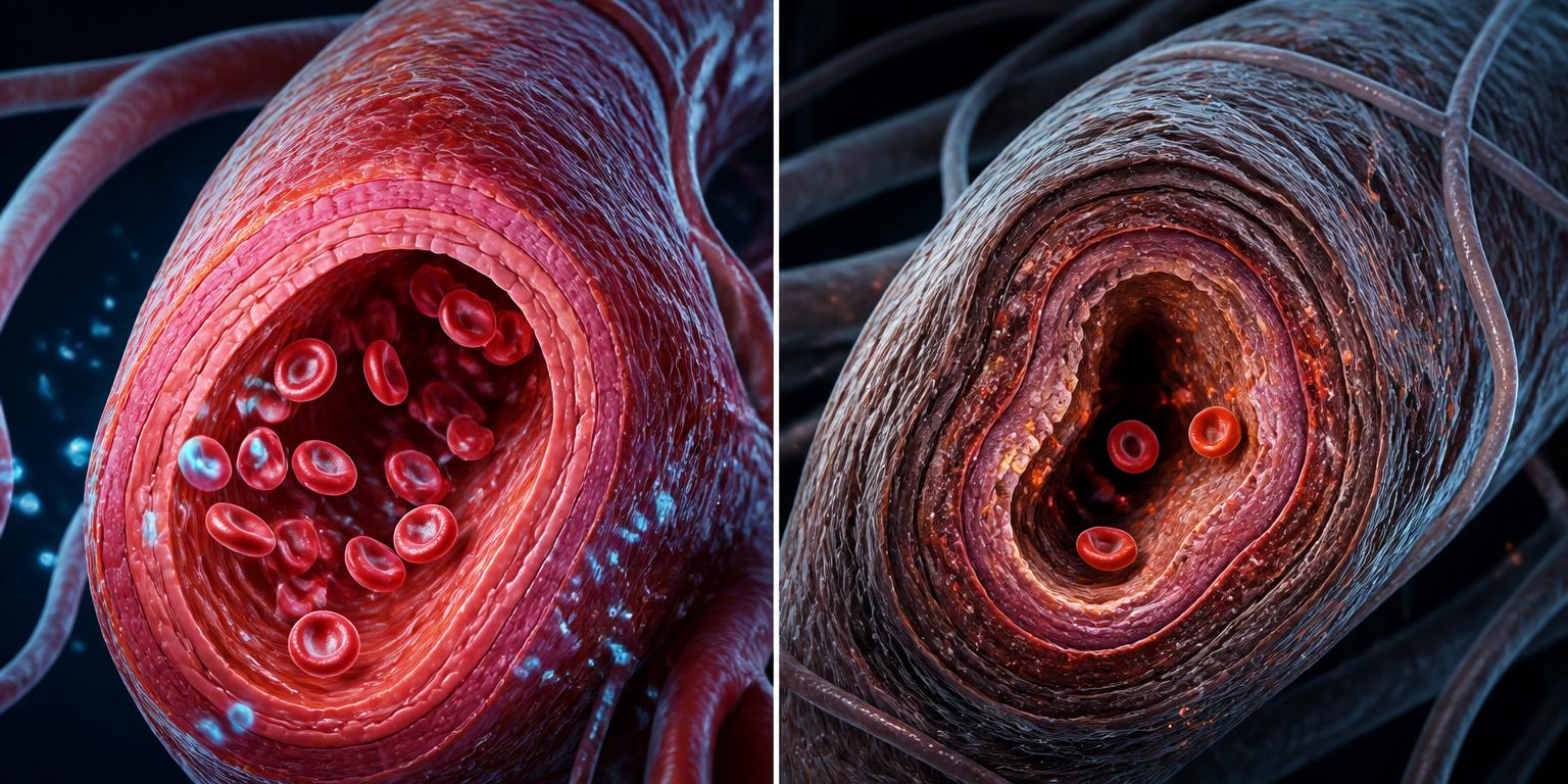
Oxidative stress is one of the most studied mechanisms in the pathophysiology of Peyronie's disease. Excessive local production of reactive oxygen species (ROS) within the injured tunica is thought to promote fibrosis, sustain low-grade inflammation and impair cavernous microcirculation.
Cascade · free radicals → plaque
Free radicals
Oxidative stress
Inflammation
Fibroblast activation
Fibrosis
Plaque
- Activation of fibroblasts and differentiation into collagen-producing myofibroblasts (fibrosis).
- Maintenance of low-grade local inflammation via NF-κB and NLRP3.
- Impairment of the cavernous microcirculation and endothelial function, contributing to associated erectile dysfunction.
Inflammatory cascade
Collagen remodelling
Microvascularisation · healthy vs dysfunctional arteriole
Consequences
- ↓ NO bioavailability
- ↑ Local oxidative stress
- ↓ Vasodilation
- ↓ Tissue oxygenation
- ↑ Fibrosis risk
To go further, see our dedicated dossiers:
Frequently asked questions
Scientific references
A selection of reference publications and international guidelines used to write this dossier. The list is not exhaustive and will be enriched as the dossier is updated.
- [1]European Association of Urology, 2024 (). EAU Guidelines on Sexual and Reproductive Health — Peyronie's disease · Voir la source
- [2]American Urological Association, 2015 (reaffirmed 2021) (). AUA Guideline: Peyronie's Disease · Voir la source
- [3]National Institute of Diabetes and Digestive and Kidney Diseases (NIDDK) (). Peyronie's Disease — Definition, Causes and Treatment · Voir la source
- [4]Loreto et al., Int J Mol Sci, 2022 (). The role of oxidative stress in Peyronie's disease · Voir la source
- [5]Safarinejad, Int J Impot Res, 2010 (). Coenzyme Q10 supplementation in patients with early chronic Peyronie's disease · Voir la source
- [8]Gonzalez-Cadavid & Rajfer, Int J Impot Res, 2005 (). Pathophysiology of Peyronie's disease · Voir la source
- [9]Joseph et al., Sex Med Rev, 2020 (). Penile traction therapy for Peyronie's disease — systematic review · Voir la source
- [10]Gelbard et al., J Urol, 2013 (). Intralesional collagenase Clostridium histolyticum for Peyronie's disease (IMPRESS trials) · Voir la source
- [11]Nelson & Mulhall, J Sex Med, 2013 (). Psychological impact of Peyronie's disease: a review · Voir la source
- [12]Raheem et al., BJU Int, 2010 (). Vacuum erection device therapy for Peyronie's disease · Voir la source
- [13]Fojecki et al., Scand J Urol, 2017 (). Low-intensity extracorporeal shockwave therapy in Peyronie's disease — meta-analysis · Voir la source
Conclusion
Peyronie's disease is a common condition, long under-diagnosed, whose management has advanced considerably over the past two decades. Current guidelines (EAU, AUA) rely on a rigorous assessment of the phase, the curvature and erectile function, with a therapeutic range from observation to corrective surgery via intralesional injections, penile traction and vacuum devices.
AARO LAB focuses on the biological mechanisms involved in oxidative stress, microvascularisation and tissue integrity. Dietary supplements do not replace medical care but can be part of an overall health approach, when their use is appropriate and discussed with a healthcare professional.
Related reading
Nitric oxide (NO)
Central regulator of vasodilation and erectile function.
Vasodilation
NO/cGMP pathway and tissue perfusion.
Cardiovascular health
Shared risk factors with erectile dysfunction.
Microcirculation
Capillary perfusion and endothelial health.
Endothelial dysfunction
Early marker involved in erectile health.
Penile fibrosis
Localised fibrosis of the tunica and cavernous bodies.
Natural approaches — fibroses
Critical reading of ingredients studied in Peyronie and Dupuytren.
Institutional sources
Further reading
These institutional resources let you explore the topic further via authoritative scientific or medical organisations.
- EAUEuropean Association of UrologyEAU Guidelines — Sexual and Reproductive HealthEuropean guidelines detailing the diagnosis and management of Peyronie's disease.
- AUAAmerican Urological AssociationAUA Guideline — Peyronie's DiseaseNorth American guideline dedicated to Peyronie's disease.
- PubMedPubMed / National Library of MedicinePeyronie's disease — clinical researchBibliographic search on epidemiology, pathophysiology and treatments.
- HASHaute Autorité de SantéHAS recommendations — urologyFrench good-practice recommendations in urology (source in French).
External links to scientific or medical organisations. AARO LAB has no commercial ties with the sites cited.